
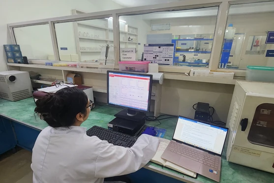
In in-vitro cell analysis, flow cytometry measures fluorescence intensity produced by labeled antibodies or ligands bound to specific cell-associated molecules — revealing what's happening on and inside a cell population without ever needing to look at a single slide under a microscope. That single measurement principle underpins an unusually wide range of applications in cultured cell work.
Core Applications in In-Vitro Cell Analysis
| Application | What It Reveals |
|---|---|
| Immunophenotyping | Identifying different cell populations based on surface markers |
| Cell Cycle Analysis | DNA content, using dyes like propidium iodide, to determine what proportion of cells are in G1, S, or G2/M phase |
| Apoptosis Detection | Annexin V/PI staining to distinguish live, early apoptotic, and dead cells |
| Intracellular Staining | Cytokines or transcription factors inside the cell, not just surface markers |
| Multiparameter Analysis | Several of the above measured simultaneously on the same sample |
Why This Matters for Cultured Cell Work
A comprehensive Flow Cytometry Full Testing Service evaluates cellular and signaling phenotypes, cell growth changes, cytokine production, cellular division, and even intracellular signaling events — all from cultured cells, without destroying the sample the way some other assays do. That combination of speed and depth is why flow cytometry sits at the center of most modern in-vitro cell analysis workflows.
Diagnostic and Research Relevance
Flow cytometry plays a crucial role in diagnostics too, offering rapid, sensitive, quantitative analysis of cells that's valuable for diagnosing and monitoring conditions including hematological malignancies (blood cancers), immunodeficiency disorders, transplantation and immune monitoring, autoimmune diseases, infectious disease, and prenatal and neonatal diagnostics.
What Flow Cytometry Data Actually Shows
- Chromosome analysis
- Cell size and quantity
- Biological processes including apoptosis and cell adhesion
- Proteomics signals relevant to early disease detection
Getting Reliable In-Vitro Cytometry Data
The same principles that make flow cytometry powerful also make it easy to get wrong: poorly designed fluorophore panels, uncontrolled autofluorescence from cultured cells, and inconsistent gating can all distort results. Standardized controls, careful panel design, and consistent sample handling are what keep in-vitro flow cytometry data comparable from one experiment to the next.
Frequently Asked Questions
What does flow cytometry measure in in-vitro cell analysis?
Flow cytometry measures fluorescence intensity produced by labeled antibodies specific to proteins on or in cells, or by ligands that bind to specific cell-associated molecules, such as propidium iodide binding to DNA.
What are the main applications of flow cytometry in cultured cells?
Core applications include immunophenotyping, cell cycle analysis, apoptosis detection, intracellular staining for cytokines or transcription factors, and multiparameter analysis combining several of these on one sample.
How is flow cytometry used in diagnostics?
Flow cytometry offers rapid, sensitive, quantitative cell analysis used in diagnosing and monitoring hematological malignancies, immunodeficiency disorders, transplantation and immune monitoring, autoimmune diseases, infectious disease, and prenatal and neonatal diagnostics.
What can go wrong with in-vitro flow cytometry data?
Poorly designed fluorophore panels, uncontrolled autofluorescence from cultured cells, and inconsistent gating can all distort results — standardized controls and consistent sample handling keep data comparable across experiments.