
Quantitative biomarker analysis turns a biological sample into a precise number, such as picograms of IL-6 per millilitre of serum. ELISA (enzyme-linked immunosorbent assay) remains the most trusted method for biomarker quantification, and modern multi-mode plate readers make it faster, more sensitive and more flexible than ever.
This guide explains how ELISA works, how multi-mode microplate readers expand detection options, and how to generate accurate, reproducible biomarker data.
What Is a Biomarker?
A biomarker is a measurable indicator of a biological state, disease or treatment response. Common examples include cytokines (IL-6, TNF-α, IL-1β), C-reactive protein (CRP), insulin, cardiac troponin, VEGF, oxidative stress markers such as 8-OHdG, and hormones such as cortisol. Quantifying biomarkers accurately supports drug discovery, preclinical studies, clinical research and diagnostics.
How ELISA Works
ELISA uses the specific binding of an antibody to its target antigen. An enzyme linked to the detection antibody, usually horseradish peroxidase (HRP) or alkaline phosphatase (ALP), converts a substrate into a measurable signal. The stronger the signal, the more biomarker in the sample.
Types of ELISA
- Direct ELISA: The antigen is coated on the plate and detected by one enzyme-labelled antibody. Fast, but less sensitive.
- Indirect ELISA: An unlabelled primary antibody plus a labelled secondary antibody amplify the signal; widely used for antibody titre testing.
- Sandwich ELISA: The gold standard for quantitative biomarker analysis. A capture antibody and a detection antibody bind two different sites on the target, giving high sensitivity and specificity.
- Competitive ELISA: Best for small molecules such as hormones and drugs; the signal falls as biomarker concentration rises.
Why Multi-Mode Plate Readers Matter
A multi-mode plate reader combines several detection technologies in one instrument. This lets one lab run classic colorimetric ELISA alongside more sensitive formats, without buying separate readers.
- Absorbance (colorimetric): Standard TMB-based ELISA read at 450 nm, with a 570 or 630 nm reference wavelength to correct for plate imperfections.
- Fluorescence intensity: Fluorogenic substrates extend sensitivity and dynamic range.
- Luminescence (chemiluminescent ELISA): Offers some of the highest sensitivity and a wide dynamic range, ideal for low-abundance biomarkers.
- Time-resolved fluorescence (TRF) and TR-FRET: Reduce background for no-wash, homogeneous assays such as HTRF.
- Fluorescence polarisation and AlphaLISA: Support high-throughput screening and wash-free biomarker detection.
Many multi-mode readers also offer temperature control, shaking, kinetic reads and 96- or 384-well plate formats, improving throughput and consistency.
From Signal to Concentration: The ELISA Standard Curve
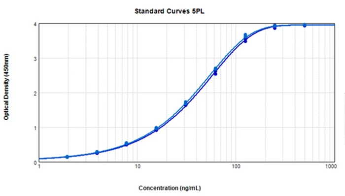
Accurate biomarker quantification depends on the standard curve. A dilution series of known standards is run on every plate, and sample concentrations are read from the fitted curve.
- Use 4PL or 5PL curve fitting: ELISA curves are sigmoidal, so four- or five-parameter logistic regression fits far better than a straight line.
- Stay within the quantifiable range: Report only values between the lower and upper limits of quantification (LLOQ and ULOQ).
- Correct for dilution: Multiply results by the sample dilution factor.
- Check fit quality: Back-calculated standards should fall close to their nominal values.
Typical Quantitative ELISA Workflow
- Coat the plate with capture antibody (or use a pre-coated ELISA kit).
- Block unbound sites to prevent non-specific binding.
- Add standards, controls and samples in duplicate or triplicate.
- Add the detection antibody, then the enzyme conjugate.
- Add substrate and stop the reaction.
- Read the plate on a microplate reader at the correct wavelength or detection mode.
- Fit the standard curve with 4PL or 5PL and calculate biomarker concentrations.
ELISA Assay Validation Essentials
Reliable biomarker data requires a validated assay. Key validation parameters include:
- Sensitivity: Limit of detection (LOD) and lower limit of quantification (LLOQ).
- Precision: Intra-assay and inter-assay coefficient of variation (CV), commonly targeted below about 10% and 15%.
- Accuracy: Spike-and-recovery, usually expected within about 80–120%.
- Dilution linearity and parallelism: Confirm that sample matrix does not distort results.
- Specificity: Check cross-reactivity with related proteins.
Best Practices for Accurate Biomarker Quantification
- Collect, process and store samples consistently, and avoid repeated freeze–thaw cycles.
- Run all standards and samples in at least duplicate.
- Watch for edge effects; equilibrate plates and reagents to room temperature.
- Dilute high samples to avoid the hook effect and to stay within the standard curve.
- Use matrix-matched standards for serum, plasma or cell culture supernatant.
- Read plates promptly after adding stop solution.
Frequently Asked Questions
Which ELISA is best for quantitative biomarker analysis?
Sandwich ELISA is the preferred format because two antibodies give high sensitivity and specificity for protein biomarkers.
What wavelength is used to read an ELISA plate?
TMB-based ELISAs are read at 450 nm after adding stop solution, often with a 570 or 630 nm reference wavelength.
What is a multi-mode plate reader?
A microplate reader that combines absorbance, fluorescence, luminescence and often advanced modes such as TRF in one instrument.
Why use 4PL curve fitting for ELISA?
ELISA standard curves are S-shaped, and four-parameter logistic fitting models that shape accurately, improving quantification at low and high concentrations.
Conclusion
ELISA combined with multi-mode plate readers is a powerful, accessible platform for quantitative biomarker analysis. Choosing the right ELISA format and detection mode, fitting standard curves correctly and validating every assay delivers sensitive, reproducible biomarker data that researchers, clinicians and regulators can trust.