
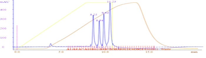
Ion exchange chromatography (IEX) separates proteins by net surface charge rather than size or affinity. Bound to a charged resin at low ionic strength, proteins are displaced in order of binding strength as a salt gradient rises — giving high capacity, high resolution, and a method that costs nothing but buffer.
pI and pH Decide Everything
A protein carries net negative charge above its isoelectric point (pI) and net positive charge below it. The resin is chosen from where the target's pI sits relative to the working buffer pH, typically set roughly one pH unit away from the pI — far enough for firm binding, close enough that contaminants of similar pI still separate.
| Mode | Resin Charge | Binds | Typical Conditions |
|---|---|---|---|
| Anion exchange (Q) | Positive | Acidic proteins | pH 8.0 Tris · protein pI < 7 |
| Cation exchange (S/SP) | Negative | Basic proteins | pH 5.5–6.5 MES/acetate · pI > 7 |
Anion exchange is the default first choice for most recombinant proteins, since typical protein pIs skew acidic. Strong exchangers (Q, S) stay fully charged across pH 2–12; weak exchangers (DEAE, CM) titrate, so their capacity shifts with buffer pH.
Buffers and Sample Conditioning
The buffering ion must carry the same charge as the resin, or it competes with the sample for binding sites — Tris (cationic) belongs on anion exchange, while acetate, MES, and phosphate (anionic) belong on cation exchange. Binding also requires low ionic strength: a sample at 150 mM NaCl will simply flow straight through a Q column, so the sample is diluted or buffer-exchanged until its conductivity matches the loading buffer.
The Run: One Method, Five Blocks
| Block | Typical Setting |
|---|---|
| Equilibrate | 5–10 CV · 0% B |
| Load | Any volume · 0% B |
| Wash | 5–10 CV · 0% B |
| Gradient elute | 10–20 CV · 0→50% B |
| Strip | 5 CV · 100% B |
Unlike affinity chromatography, IEX load volume is effectively unlimited — the protein concentrates on the column as it binds, so a dilute sample is actually an advantage.
Gradient Shape Sets Resolution
Proteins elute when rising salt concentration overcomes their electrostatic grip on the resin, so gradient steepness (percent B per column volume) is the single most powerful resolution control. Shallow gradients separate closely related species; steep gradients concentrate. A common strategy is to scout broad first (0→100% B over 20 CV) to find where the target elutes, then narrow the gradient around that point for optimal resolution.
How Do You Read an Ion Exchange Chromatogram?
| Observation | Interpretation |
|---|---|
| Everything in flow-through | Wrong resin charge, wrong pH, or sample conductivity too high |
| Peak only on 100% strip | Binding too strong — raise pH toward the pI or extend the gradient |
| High A260 relative to A280 | Nucleic acid bound to the resin — common on anion exchange from crude lysate |
Frequently Asked Questions
Why did nothing bind to my column?
The most common cause is a sample that is too salty for the resin to bind it — dilute or buffer-exchange the sample until its conductivity matches the loading buffer.
Should I record the elution fraction number or the conductivity?
Always record the elution conductivity or %B, not the fraction number — conductivity transfers between columns and systems, while fraction volume does not.
Conclusion
A well-designed IEX method comes down to matching resin charge and buffer pH to your target's pI, conditioning the sample to low ionic strength, and choosing a gradient shape suited to your resolution needs. Our protein purification team can help design and troubleshoot IEX steps for both research and process-scale work.