
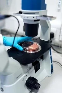
Microscopy is how cell biology becomes visible. Different microscopy techniques reveal different layers of information — from basic morphology to the precise location of a single protein inside a cell — and choosing the right one depends entirely on the question being asked.
Brightfield and phase-contrast microscopy
These are the everyday workhorses of a cell culture lab. Brightfield microscopy is used to check confluency, monitor morphology, and spot contamination during routine culture maintenance. Phase-contrast enhances the visibility of unstained, transparent cells by exploiting differences in refractive index, which makes it much easier to assess live cell health without needing to fix or stain the sample.
Fluorescence microscopy
Fluorescence microscopy uses fluorescently tagged antibodies, dyes, or proteins to visualize specific structures or molecules within a cell. It's the basis for immunofluorescence staining, where an antibody-dye conjugate binds to a target protein, revealing its subcellular location — nucleus, cytoplasm, membrane, or specific organelles — under UV or laser excitation.
Immunofluorescence in practice
- Protein localization — confirming where a protein of interest sits within the cell
- Co-localization studies — using multiple fluorophores to see whether two proteins occupy the same cellular compartment
- Pathology and diagnostics — identifying disease markers in tissue sections
- Cytoskeletal and structural studies — visualizing actin, tubulin, or other structural proteins
Histology and tissue-level microscopy
For tissue samples rather than cultured cells, histological staining combined with microscopy reveals structural and pathological detail — this is central to diagnosing disease from biopsy samples and to studying tissue architecture in research models.
How Do You Choose the Right Microscopy Technique?
A quick confluency check doesn't need fluorescence; a co-localization study can't be done without it. Matching the microscopy method to the actual biological question — and preparing the sample correctly for that method, whether that's fixation, permeabilization, or antigen retrieval — is what determines whether the resulting images actually answer the question at hand.
Our lab offers immunofluorescence microscopy services and a hands-on workshop covering tissue preparation, staining, and image-based analysis.
Frequently Asked Questions
When should I use fluorescence microscopy instead of brightfield?
Brightfield and phase-contrast are the everyday tools for checking confluency, morphology, and contamination during routine culture maintenance. Fluorescence microscopy is needed when you must visualize specific structures or molecules -- such as confirming a protein's subcellular location -- using fluorescently tagged antibodies, dyes, or proteins.
What sample preparation does immunofluorescence typically require?
Depending on the target, immunofluorescence sample preparation may involve fixation, permeabilization, or antigen retrieval -- matching the preparation to the biological question is what determines whether the resulting images actually answer it.