
Apoptosis, or programmed cell death, is central to cancer research, drug discovery, toxicology and immunology. Among the many apoptosis assays available, Annexin V / PI staining analysed by flow cytometry is the most widely used. It is fast, quantitative and, crucially, separates live, early apoptotic, late apoptotic and necrotic cells in a single sample.
This guide explains the principle of the Annexin V apoptosis assay, a step-by-step protocol, how to read the results and how to avoid the mistakes that lead to false positives.
The Principle of Annexin V / PI Staining
Annexin V Detects Phosphatidylserine
In healthy cells, phosphatidylserine (PS) is kept on the inner side of the plasma membrane. Early in apoptosis, PS flips to the outer surface, where it acts as an "eat me" signal for phagocytes. Annexin V is a protein that binds PS strongly in the presence of calcium. Labelled with a fluorochrome such as FITC, PE or APC, Annexin V marks cells that have exposed PS.
Propidium Iodide Detects Membrane Damage
Propidium iodide (PI) is a DNA-binding dye that cannot cross an intact cell membrane. It only stains cells whose membranes are compromised, as in late apoptosis and necrosis. 7-AAD is a common alternative to PI.
Because Annexin V can also reach PS on the inner membrane of damaged cells, PI is needed to tell early apoptosis apart from late apoptosis and necrosis.
How to Interpret Annexin V / PI Results
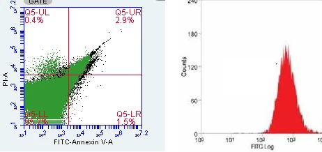
Results are displayed as a dot plot of Annexin V against PI, divided into four quadrants:
- Annexin V negative / PI negative: Live, healthy cells.
- Annexin V positive / PI negative: Early apoptotic cells, with PS exposed but membranes still intact.
- Annexin V positive / PI positive: Late apoptotic or secondary necrotic cells.
- Annexin V negative / PI positive: Necrotic cells or damaged nuclei and debris.
Total apoptosis is usually reported as early plus late apoptotic cells. Running a time course helps show the progression from early to late apoptosis.
Annexin V / PI Staining Protocol
- Treat cells with the test compound alongside untreated and positive controls (for example, staurosporine or camptothecin).
- Collect all cells, including floating cells in the medium, since many apoptotic cells detach.
- Detach adherent cells gently, for example with Accutase or brief, mild trypsinisation, to avoid damaging membranes.
- Wash cells in cold PBS and resuspend in calcium-containing Annexin V binding buffer at about 1 × 10⁶ cells/mL.
- Add fluorescent Annexin V and PI, and incubate for about 15 minutes at room temperature in the dark.
- Add more binding buffer and analyse by flow cytometry as soon as possible, ideally within one hour.
Do not fix cells before Annexin V / PI staining: fixation permeabilises membranes and makes every cell PI-positive.
Essential Controls
- Unstained cells: Set voltages and measure autofluorescence.
- Annexin V-only and PI-only controls: Needed for compensation, since FITC and PI emissions overlap.
- Positive apoptosis control: Cells treated with a known apoptosis inducer confirm the assay works.
- Untreated or vehicle control: Defines baseline apoptosis in the culture.
Flow Cytometry Gating Tips
- Gate on cells using FSC and SSC, but keep the gate wide: apoptotic cells shrink and become more granular, so a tight gate will exclude them.
- Exclude doublets with an FSC-A vs FSC-H plot.
- Set quadrant boundaries using the single-stained and unstained controls.
- Collect enough events, typically at least 10,000 cells per sample, for reliable percentages.
Common Mistakes and How to Avoid Them
- Harsh cell harvesting: Over-trypsinisation or scraping damages membranes and creates false PI-positive and Annexin V-positive cells.
- Using EDTA or calcium-free buffers: Annexin V binding needs calcium; EDTA chelates it and reduces staining.
- Discarding floating cells: This removes many apoptotic cells and underestimates cell death.
- Delays before analysis: Early apoptotic cells progress to late apoptosis if left too long.
- Fluorescent proteins or drugs: GFP-expressing cells or fluorescent compounds need a different fluorochrome, such as Annexin V-APC with 7-AAD.
- Poor compensation: Uncompensated FITC/PI spillover shifts cells into the wrong quadrant.
Confirming Apoptosis With Complementary Assays
Annexin V / PI staining is powerful, but combining it with another method strengthens conclusions:
- Caspase-3/7 activity assays: Confirm activation of the apoptotic pathway.
- Mitochondrial membrane potential (JC-1 or TMRM): Detects early mitochondrial changes in intrinsic apoptosis.
- TUNEL assay: Detects DNA fragmentation in cells and tissue sections.
- Western blotting: Shows cleaved caspase-3, cleaved PARP and Bcl-2 family proteins.
- Cell cycle analysis: A sub-G1 peak indicates fragmented DNA in apoptotic cells.
Frequently Asked Questions
What does Annexin V / PI staining detect?
It detects apoptosis by identifying phosphatidylserine exposure (Annexin V) and loss of membrane integrity (PI), separating live, early apoptotic, late apoptotic and necrotic cells.
What is the difference between early and late apoptosis?
Early apoptotic cells are Annexin V-positive and PI-negative with intact membranes; late apoptotic cells are positive for both, as their membranes have become permeable.
Why does Annexin V need calcium?
Annexin V binds phosphatidylserine only in the presence of calcium ions, so a calcium-containing binding buffer is essential.
Can Annexin V / PI stained cells be fixed?
Not before staining. Fixation permeabilises membranes and causes false PI staining. Analyse fresh cells promptly for the most accurate results.
Conclusion
Annexin V / PI staining is the go-to apoptosis assay for quantifying early and late apoptosis by flow cytometry. Gentle cell handling, calcium-containing buffer, proper compensation controls and prompt analysis are the keys to accurate results, and pairing it with caspase, mitochondrial or DNA fragmentation assays provides strong, publication-ready evidence of apoptotic cell death.