
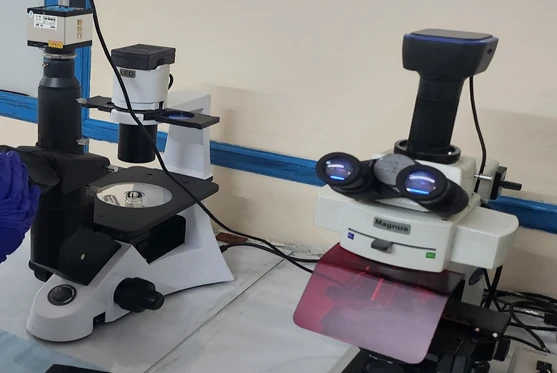
Before any antibody is added, two channels already describe a cell on a flow cytometer. Forward scatter (FSC) tracks how much light a cell deflects at small angles — broadly its size — while side scatter (SSC) tracks light refracted at 90° by internal structure — its granularity and nuclear complexity. A scatter plot is not an image, but across a population it can separate what microscopy separates, at thousands of cells per second.
Two Angles, Two Properties
Forward scatter is light collected within a few degrees of the beam axis. It scales with a cell's cross-sectional area and the refractive-index difference between the cell and its medium, so it correlates with size but is not a calibrated diameter — it should be read as relative size, never as a µm measurement. Side scatter is light refracted and reflected at right angles by internal interfaces: granules, lysosomes, mitochondria, vesicles, and nuclear lobes. Densely granular cells scatter far more light than smooth cells of the same diameter.
How Do You Read the FSC/SSC Plane?
| Position | Typical Population |
|---|---|
| Low FSC, high SSC | Small and complex — apoptotic bodies, degranulating fragments, some bacteria and platelets |
| High FSC, high SSC | Large and granular — granulocytes, activated macrophages, lipid-laden cells |
| Low FSC, low SSC | Debris zone — fragments and free nuclei, though shrunken apoptotic cells drift here too |
| High FSC, low SSC | Large and smooth — lymphoblasts, most cultured lines, cells in G2/M |
Pulse Geometry: The Third Dimension
Each event is a pulse with shape as well as amplitude. Height is peak brightness, area is the integrated signal, and width is transit time through the beam — a proxy for how long the object is along the flow axis. A doublet has roughly double the area of a singlet but a similar height, which is exactly why FSC-A plotted against FSC-H separates doublets cleanly while FSC-H alone cannot.
Preparation Changes Morphology
Scatter is the parameter most sensitive to handling. Trypsin rounds cells and strips surface projections; fixation shrinks cells and raises SSC; osmotic stress swells or crenates them. Every condition in a comparison series needs to be prepared identically and to the same timing, or the morphology "difference" measured is really just the difference in preparation.
Anchoring the Axes
Scatter has no natural units, so a series is only comparable if something fixed appears in every tube. Calibration beads of known diameter give an approximate size ladder (beads refract light differently from cells, so this is a ladder, not a true calibration), while a stable internal reference population — unstimulated cells expected not to change — is often the more reliable anchor in practice.
Morphological Signatures Worth Reading
| State | Scatter Shift | Confirm With |
|---|---|---|
| Early apoptosis | FSC ↓, SSC ↑ then ↓ | Annexin V |
| Necrosis | FSC ↓↓, SSC ↓ | PI / 7-AAD |
| Blast activation | FSC ↑, SSC modest ↑ | CD69 / CD25 |
Scatter suggests a cell state; it does not establish one on its own. Every signature above needs its confirming marker before the claim is made.
Frequently Asked Questions
Can I compare scatter values between two different instruments?
Not directly. Optics and detector gain differ between instruments, so scatter values do not transfer without a common anchor such as calibration beads run on both machines.
Why does a treated sample appear to have "no effect" on morphology?
A debris gate that is drawn too tightly can exclude shrunken apoptotic cells, making a real treatment effect disappear. Set the gate generously and confirm with a lineage or viability marker.
Conclusion
Scatter-based morphology analysis is a fast, label-free first look at a population, but it always needs a fixed reference and a confirming marker before a biological claim is made. Our cell biology and flow cytometry services include full morphological and viability characterization.