
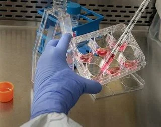
Flow cytometry measures physical and chemical properties of individual cells as they flow one by one through a laser beam. Because it looks at cells one at a time rather than as a bulk population, it generates detailed, multi-parameter data on thousands of individual cells in minutes — making it a core technique in immunology, oncology, and molecular research.
What Flow Cytometry Actually Measures
As each cell passes through the laser, forward scatter and side scatter reveal its size and internal complexity, while fluorescent antibodies or dyes bound to specific markers reveal what proteins are expressed on or inside the cell. Combining several fluorescent channels together makes it possible to distinguish cell types, activation states, and viability all from a single sample run.
Why It Matters in Research
Single-Cell Resolution at Scale
Thousands of individual cells per second are analyzed, providing statistically robust data on heterogeneous populations.
Multiparametric Analysis
Modern instruments detect 10 to over 40 parameters simultaneously, allowing deep characterization of complex cell subsets in one run.
Cell Sorting Capability
Fluorescence-Activated Cell Sorting (FACS) physically separates live cells based on chosen markers for downstream culture or molecular analysis.
High-Throughput Quantitative Data
Flow cytometry generates statistically powerful, quantitative data rapidly, supporting large-scale immunology and clinical research studies.
Broad Research Relevance
The technique is central to immunology, hematology, stem cell research, and oncology.
Common Uses
- Immunophenotyping — identifying and counting specific immune cell populations (T cells, B cells, NK cells) based on surface markers
- Cell cycle analysis — using DNA-binding dyes like propidium iodide to determine what proportion of cells are in G1, S, or G2/M phase
- Apoptosis detection — Annexin V/PI staining distinguishes live, early apoptotic, and dead cells
- Clinical diagnostics — flow-based FACS panels are routine in leukemia and lymphoma diagnosis and treatment monitoring
- Drug and toxicity screening — assessing how a compound affects cell viability at the single-cell level
Flow Cytometry vs. Fluorescence Microscopy
| Feature | Flow Cytometry | Fluorescence Microscopy |
|---|---|---|
| Throughput | Thousands of cells per second | Limited to field of view |
| Quantification | Highly quantitative, statistical | Qualitative to semi-quantitative |
| Cell Sorting | Possible (FACS) | Not possible |
| Parameters per Cell | Up to 40+ simultaneously | Typically fewer, filter-limited |
Why Single-Cell Resolution Matters
- Bulk assays report an average across an entire population, which can hide important biology
- A rare subpopulation of resistant cells would be invisible in a bulk measurement but clearly visible in flow cytometry data
- This resolution is especially valuable in oncology and immunology research
- It supports detection of minimal residual disease and rare-event analysis
Getting Reliable Results
- Ensure proper instrument setup and calibration using standardized beads
- Design fluorophore panels thoughtfully to avoid spillover
- Use viability dyes to exclude dead cells from analysis
- Apply consistent, well-controlled gating strategies based on defined controls (FMOs)
Frequently Asked Questions
What is the difference between flow cytometry and FACS?
Flow cytometry refers broadly to the analysis of cells in a fluid stream, while FACS (Fluorescence-Activated Cell Sorting) is a specific application that also physically sorts cells based on selected markers.
What is gating in flow cytometry?
Gating is the process of selecting a specific population of cells within flow cytometry data, based on scatter or fluorescence properties, to focus analysis on a subset of interest.
How many parameters can flow cytometry measure at once?
Modern flow cytometers can measure over 40 parameters simultaneously, depending on the number of lasers and detectors available.
What sample types can be used for flow cytometry?
Flow cytometry requires single-cell suspensions, which can be prepared from blood, dissociated tissue, or cultured cells.
Conclusion
Good flow cytometry data depends on proper instrument setup and calibration, thoughtful panel design, and consistent sample preparation. Our clinical FACS and flow cytometry services cover instrument set-up, experimental design, and full data analysis and interpretation support.