
An oxidative stress assay measures the balance between reactive oxygen species (ROS) and the body's antioxidant defences. In-vitro oxidative stress assays are the fastest, most cost-effective way to screen drugs, plant extracts, nutraceuticals and cosmetics for antioxidant activity, or to flag toxic compounds that trigger oxidative damage, before moving to animal or clinical studies.
A reliable study measures three things: ROS levels, oxidative damage, and antioxidant defence. Here are the key assays for each.
What Is Oxidative Stress?
Oxidative stress occurs when free radicals such as superoxide, hydrogen peroxide (H₂O₂) and hydroxyl radicals overwhelm antioxidant defences. The result is damage to lipids, proteins and DNA, linked to ageing, diabetes, cancer, neurodegeneration and cardiovascular disease. Measuring oxidative stress markers in vitro shows whether a test compound prevents or causes this damage.
1. Chemical Antioxidant Assays (Cell-Free)
These quick, low-cost screening tests rank the free radical scavenging activity of a sample.
- DPPH assay: The most popular antioxidant assay. Antioxidants reduce the purple DPPH radical, lowering absorbance at 517 nm. Results are reported as % inhibition and IC50 value.
- ABTS assay: Measures decolourisation of the ABTS radical cation at 734 nm; works for both water- and fat-soluble antioxidants. Reported as Trolox equivalents (TEAC).
- FRAP assay: Ferric reducing antioxidant power; measures reduction of Fe³⁺ to Fe²⁺ at 593 nm.
- ORAC assay: Oxygen radical absorbance capacity; tracks protection of a fluorescent probe from peroxyl radicals.
- Radical scavenging assays: Hydroxyl radical, superoxide and nitric oxide scavenging tests add mechanism-specific evidence.
Tip: run at least two assays (e.g., DPPH and FRAP), since each works by a different mechanism.
2. Cellular ROS Assays
Cell-based assays confirm antioxidant activity inside living cells, which is far more meaningful than chemistry alone. Common cell lines include HepG2, HaCaT, SH-SY5Y, PC12 and RAW 264.7.
- DCFH-DA assay: The standard intracellular ROS assay. Cells oxidise the probe to fluorescent DCF, read by plate reader, flow cytometry or microscopy.
- MitoSOX / DHE: Detects superoxide, including mitochondrial ROS.
- Amplex Red: Sensitive detection of hydrogen peroxide released by cells.
3. Oxidative Damage Markers
- MDA / TBARS assay: The most widely used lipid peroxidation assay, measuring malondialdehyde (MDA) at 532 nm.
- 4-HNE: A more specific lipid peroxidation marker, measured by ELISA.
- Protein carbonyl assay: Detects protein oxidation using DNPH.
- 8-OHdG assay: The key marker of oxidative DNA damage, measured by ELISA or LC-MS/MS.
4. Antioxidant Enzyme Assays
These tests show whether a compound strengthens the cell's own defences.
- SOD assay: Superoxide dismutase activity.
- Catalase assay: Breakdown of H₂O₂, read at 240 nm.
- GPx assay: Glutathione peroxidase activity via NADPH consumption.
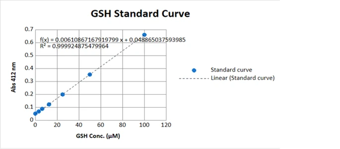
- GSH and GSH/GSSG ratio: Reduced glutathione level; a falling ratio is an early sign of oxidative stress.
- Nrf2 pathway: Nrf2, HO-1 and NQO1 expression reveal the mechanism behind protection.
Typical In-Vitro Oxidative Stress Study Design
- Screen antioxidant capacity with DPPH, ABTS or FRAP.
- Find a non-toxic dose using an MTT cell viability assay.
- Pre-treat cells with the test compound.
- Induce oxidative stress with H₂O₂, t-BHP or another inducer.
- Measure ROS (DCFH-DA), damage (MDA) and defences (SOD, CAT, GSH).
- Compare against a positive control such as N-acetylcysteine (NAC) or ascorbic acid.
Best Practices for Accurate Results
- Combine several assays; never rely on DPPH alone.
- Run sample blanks for coloured plant extracts.
- Normalise results to protein content or cell number.
- Include untreated, stressor-only, vehicle and positive controls.
- Use at least three independent experiments.
Frequently Asked Questions
What is the best assay for oxidative stress?
No single assay is enough. A strong panel combines DPPH or FRAP (antioxidant capacity), DCFH-DA (ROS), MDA (lipid peroxidation) and SOD, catalase or GSH (antioxidant defence).
What is the difference between the DPPH and ABTS assays?
DPPH works mainly in organic solvents and suits lipophilic antioxidants; ABTS works in both aqueous and organic systems, so it measures a wider range of compounds.
Which marker is used for lipid peroxidation?
Malondialdehyde (MDA), measured by the TBARS assay, is the most common lipid peroxidation marker.
How is oxidative stress induced in vitro?
Usually by exposing cells to hydrogen peroxide (H₂O₂), tert-butyl hydroperoxide, menadione or high glucose.
Conclusion
In-vitro oxidative stress assays give a fast, ethical and reliable picture of a compound's antioxidant potential. Pairing antioxidant assays like DPPH and FRAP with cellular ROS, MDA and antioxidant enzyme assays delivers publication-ready, mechanism-based evidence for pharmaceutical, nutraceutical, herbal and cosmetic research.